A broken bone does not become strong again the moment pain fades or an X-ray shows new bone. Recovery depends on the biology of repair, the fracture’s stability and blood supply, the treatment used, and how carefully movement is reintroduced. By the end, you will know what changes to expect, how clinicians judge progress, and which problems require prompt review.
Key takeaways
- Expect inflammation, soft callus, hard callus and bone remodelling.
- Healing time depends on the bone, fracture pattern, blood supply and your health.
- Seek urgent care for numbness, pale or cold skin, severe swelling or an open wound.
- Follow prescribed weight limits and rehabilitation exercises before increasing activity.
What happens inside the bone after a fracture?
A broken bone does not knit back in one instant. The fracture healing stages move from bleeding and inflammation to temporary bridging, new bone formation and gradual reshaping. What you feel and what an X-ray shows can differ during each stage.
- Inflammation after fracture begins immediately. Torn blood vessels fill the gap with a fracture haematoma, while swelling, bruising, warmth and tenderness develop around the injury. Movement is painful because nearby muscles and soft tissues are also damaged.
- During the soft-callus phase, fibrous tissue and cartilage grow across the break. This soft callus reduces movement between the fragments, but the area remains weak and normal loading can disrupt it.
- During the hard-callus phase, immature bone gradually replaces the soft callus. Stability improves, and pain with gentle movement often decreases. Visible callus is new repair tissue, not proof that the bone has regained its original strength.
- Remodelling continues after the fracture feels more secure. Bone is slowly reshaped along lines of stress, becoming more organised and stronger over time.
Pain can persist after the fracture is stable because swelling, muscle weakness, soft-tissue injury and joint stiffness take longer to settle. Reduced pain alone does not prove that normal loading is safe; examination and prescribed follow-up imaging determine when activity can increase.
How long does healing take, and how is progress checked?
Six weeks is a rough expectation for some uncomplicated injuries, not a universal deadline. The six weeks fracture myth ignores the bone involved, fracture pattern and the person’s biology.
| Bone or injury | Usual pattern |
|---|---|
| Wrist fracture | An uncomplicated, well-aligned fracture may become sufficiently stable in several weeks; stiffness and weakness can last longer. |
| Ankle fracture | Healing often takes several weeks to months, especially when the joint surface is involved or weight-bearing is restricted. |
| Femur fracture | This large, heavily loaded bone commonly needs months, particularly after severe displacement or extensive soft-tissue injury. |
| Vertebral fracture | A stable compression fracture may improve over weeks to months; instability, nerve symptoms or multiple fractures change the plan. |
Fracture healing time also changes with displacement, a gap between fragments, blood supply, age, general health and treatment. A stable fracture in a younger person may progress quickly; a severe injury with damaged tissue or poor circulation can take much longer.
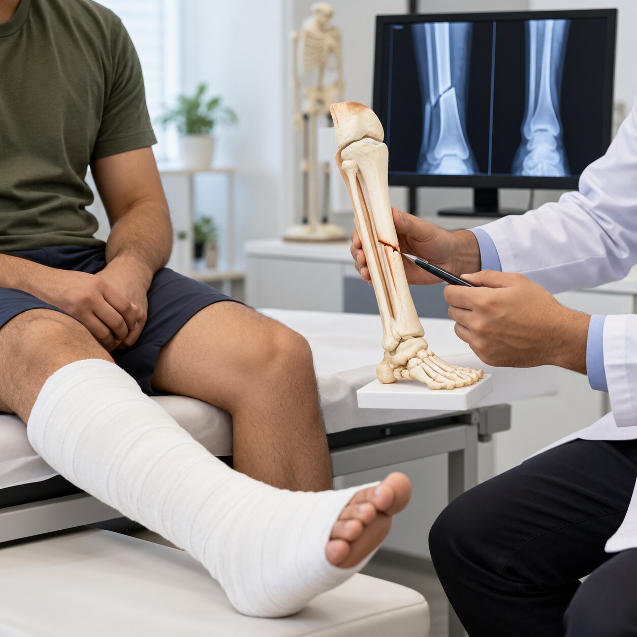
Clinical healing means examination shows less focal tenderness, improved alignment and stability, reduced swelling, better movement and safe progression of loading. Radiographic healing means follow-up X-rays show bridging bone across the fracture. Neither pain relief nor a visible X-ray change proves the other has occurred.
Clinicians interpret both findings together. CT helps define complex fracture lines and joint surfaces; MRI is useful for selected questions about soft tissue, bone marrow or an occult injury. A fracture can remain painful after becoming stable, while reduced pain alone does not make normal loading safe.
Which treatment gives a fracture enough stability to heal?
The right treatment provides enough stability for bone ends to stay aligned while healing, without immobilising more than necessary. Comparing a fracture cast versus splint helps: a splint accommodates early swelling, while a cast restricts movement more fully.
| Option | What it does | Main limit or use |
|---|---|---|
| Splint | Supports the limb while leaving room for swelling | Useful early, but allows more movement |
| Cast | Restricts movement around the fracture | Tightening swelling can affect circulation |
| Brace | Provides controlled support and movement | Works only when alignment is stable |
| Traction | Uses steady pulling force to maintain position | Selected injuries and temporary stabilisation |
| Closed reduction | Realigns fragments without an incision | Alignment can be lost afterward |
| Internal fixation plates and screws | Holds alignment inside the body | Requires surgery and hardware can cause problems |
| External fixation | Pins connect the bone to a frame outside the body | Pin-site infection and limited comfort |
Non-operative care is reasonable when fragments remain acceptably aligned, the fracture is stable and the skin, nerves and blood vessels are safe. Surgery becomes more likely with:
- An open fracture or severe soft-tissue damage
- Unstable or displaced fragments
- Joint-surface involvement
- Nerve or blood-vessel injury
- Failure to maintain acceptable alignment
Surgery restores alignment and stability; it does not automatically speed biological healing. Risks include infection, stiffness, nerve or vessel injury and hardware problems. In Panvel, Dhanvantari Hospital can help patients discuss these trade-offs and follow-up criteria with an orthopaedic team.
Related services
Fractures Treatment & Surgery At Dhanvantari Hospital, Dr. Anand Upadhye and Dr. Sushant Upadhye provides expert diagnosis and advanced treatment for all types of bone fractures,... View service → |
Accident and Trauma Surgery At Dhanvantari Hospital, Dr. Anand Upadhye and Dr. Sushant Upadhye provides expert Accident and Trauma Surgery treatment for patients suffering from... View service → |
What can delay healing or turn a fracture into an emergency?
Poor alignment, excessive movement or a gap between fragments can prevent a stable bridge of bone from forming. Reduced blood supply, infection and severe soft-tissue damage add biological obstacles. Smoking and fracture healing are closely linked: nicotine reduces blood flow and interferes with bone-forming cells.
Delayed union means healing is taking longer than expected. A nonunion fracture has failed to progress toward healing. Diabetes, poor nutrition and some medicines can also affect repair, so clinicians may review your health, medication list and X-rays before choosing treatment.
Treatment depends on the cause. Options include improving fixation, correcting alignment, treating infection, bone grafting or revision surgery. Surgery restores stability; it does not automatically make bone heal faster.
An open fracture emergency requires immediate care, even when the wound looks small. Cover it with a clean dressing, do not repeatedly explore or wash inside it, and seek emergency treatment for intravenous antibiotics and surgical cleaning and removal of contaminated or dead tissue.
NSAIDs are not automatically forbidden after a fracture. Clinicians weigh their pain-control benefit against kidney disease, ulcer bleeding, anticoagulants and an unusually high-risk fracture; ask before using them rather than stopping prescribed pain relief yourself.
Seek urgent medical review for:
- Numbness or a new loss of movement
- A pale or cold limb
- Worsening swelling or uncontrolled pain
- Fever or wound drainage
Smoking cessation during treatment is a meaningful part of fracture care, not merely general-health advice. Any worsening symptom deserves prompt reassessment, because a problem with circulation, infection or pressure can threaten the limb.
How should movement, rehabilitation and future fracture prevention be managed?
Rehabilitation can begin before an X-ray shows complete healing when the fracture and fixation are stable enough. Immobilisation causes loss of joint motion, muscle strength and balance, so fracture rehabilitation progresses in stages rather than waiting for perfect images.
| Stage | Main work | Safety rule |
|---|---|---|
| Early, protected | Gentle range-of-motion exercises for permitted joints | Keep the fracture and fixation within prescribed limits |
| Progressive | Muscle strengthening and balance training | Stop if pain, swelling or instability increases |
| Loading phase | Gradual weight-bearing after fracture and return to function | Advance only at the clinician’s stated pace |
Prescribed restrictions take priority. Loading too soon can shift fragments or loosen screws, while pushing through pain or “testing” the limb can jeopardise alignment; less pain does not prove that normal loading is safe.
Children with a growth-plate injury need growth plate fracture follow-up even after an early X-ray looks satisfactory. Monitoring checks for growth arrest, limb-length difference and angular deformity.
For an older adult with a low-energy fracture, osteoporosis after fracture deserves active planning. Ask about bone-density and falls assessment, calcium and vitamin D advice, safe exercise, home hazards and bone-strengthening medicine.
At each follow-up, ask:
- Is alignment maintained?
- What movement and loading are allowed now?
- When is the next X-ray?
- Which symptoms mean I should return sooner?
Frequently asked questions
What happens inside the bone after a fracture?
Blood collects at the break, inflammation begins, a soft callus bridges the gap, hard bone forms, and remodelling strengthens the repaired area.
How long does a fracture take to heal?
Healing time varies by the bone, fracture type, blood supply, age, smoking, medical conditions and treatment. X-rays and examination track progress.
How is fracture healing progress checked?
A clinician checks pain, tenderness, swelling, movement and function, then compares follow-up X-rays for bridging bone and alignment.
When does a fracture become an emergency?
Get urgent care for an open wound, uncontrolled bleeding, numbness, loss of movement, pale or blue skin, a cold limb, severe deformity or rapidly increasing pain.
How can you support recovery and prevent future fractures?
Use the prescribed splint or cast, follow weight-bearing limits, complete rehabilitation, eat adequately, stop smoking and address fall and bone-health risks.
Related service
Paediatric Orthopedics Treatment Paediatric Orthopedics Treatment Dr. Anand Upadhye and Dr. Sushant Upadhye at Dhanvantari Hospital provides specialized paediatric orthopedics care for children with bone, joint,... View service → |