Joint pain does not automatically mean that a camera examination or surgery will help. By understanding what arthroscopy can reveal, what it can treat and how its risks compare with MRI, physiotherapy and other options, you can have a more useful discussion about the next step.
Key takeaways
- An arthroscope uses a camera, light and saline to inspect a joint through small portals.
- Surgeons can repair selected cartilage, ligament, meniscus and synovial problems during arthroscopy.
- Recovery time depends on the tissue repaired, not the size of the skin incisions.
- Ask whether physiotherapy, injections or open surgery offers a better result for your problem.
What the arthroscope shows inside a joint
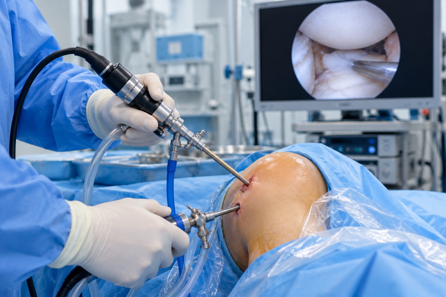
An arthroscope is a narrow camera with a light source that enters the joint through a small portal. Saline irrigation gently expands the space and clears blood or debris, while separate arthroscopy portals allow miniature instruments to inspect or treat tissue.
| Joint | What direct inspection can show |
|---|---|
| Knee | Cartilage, menisci, ligaments, synovium and loose bone or cartilage bodies |
| Shoulder | Labrum, rotator-cuff-related structures, cartilage, biceps-related tissue and loose bodies |
| Hip | Labrum, cartilage, synovium, impingement-related damage and loose bodies |
| Ankle | Cartilage, synovium, ligaments and loose fragments |
| Wrist | Cartilage, ligaments, synovium and selected triangular fibrocartilage injuries |
| Elbow | Cartilage, synovium, ligaments and loose bodies |
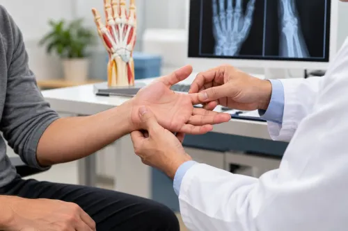
A camera view is evidence, not a diagnosis by itself. The surgeon compares what the arthroscope shows with your symptoms, physical examination and X-rays or MRI because the suspected tear may not be the true pain source. A normal-looking joint can redirect investigation toward referred pain, tendon problems, or nerve and spine causes.
Ask about knee arthroscopy diagnosis and treatment experience of pain practitioners and orthopaedic surgeons involved in your care. Direct findings can support treatment during the same procedure, but arthroscopy remains surgery, even when the portals are small.
What can be treated during the same procedure
Diagnostic arthroscopy inspects the joint and may take fluid or synovial tissue for testing; therapeutic arthroscopy treats a suitable finding during the same operation, with consent for that possibility. Treatment can include removing loose bodies, synovitis treatment, smoothing or selectively removing damaged tissue, and repairing suitable meniscal or labral tears.
| Meniscus repair versus meniscectomy | Benefit | Trade-off |
|---|---|---|
| Repair | Preserves load-distributing tissue and limits added joint stress | Slower rehabilitation; the repair can fail to heal |
| Partial meniscectomy | Often allows faster early weight bearing and recovery | Removes tissue and increases contact stress in the joint |
Selected ligament procedures are also possible. ACL reconstruction is not simply removal of a torn ligament: the surgeon creates bone tunnels and passes a tendon graft through them, usually with arthroscopy assistance in arthroscopic surgery.
Shoulder procedures can address selected rotator-cuff tears, labral tears, instability, biceps problems and some cartilage defects. Arthroscopy cannot restore lost articular cartilage, reverse established osteoarthritis or guarantee pain relief. Small portals do not make surgery risk-free; anaesthesia, infection, bleeding, clots, stiffness, nerve injury and persistent pain remain possible.
When arthroscopy is the right choice—and when another option is better
Arthroscopy is the right choice when your symptoms, examination and imaging point to a specific structural problem that the surgeon can treat, and the result matches your activity goals. MRI can show a tear, but it cannot decide whether surgery will improve pain or function.
Plain radiographs show alignment, fractures and arthritis that MRI may not answer. Clinicians also compare symptom patterns with examination findings: a degenerative meniscus tear is not automatically an indication for surgery, and intermittent clicking does not prove that arthroscopy will help.
| Option | What it can do | Important boundary |
|---|---|---|
| Arthroscopy versus MRI | Inspect and treat a suspected lesion directly | MRI shows anatomy but does not predict surgical benefit |
| Arthroscopy versus physiotherapy | Address a repairable structural problem | Physiotherapy improves strength and movement without surgical risk |
| Injection | Test a pain source or reduce inflammation | It does not repair torn tissue |
| Open surgery | Reach or reconstruct work that portals cannot complete | Larger exposure may be necessary |
Do not routinely use knee lavage or debridement for osteoarthritis. A displaced fragment that physically prevents motion is different: the phrase true locked knee arthroscopy indications describes this mechanical problem, not occasional catching.
Advanced hip arthritis, infection, fracture, major deformity or diffuse pain without a structural cause require another plan. That may include medication, rehabilitation or joint replacement when arthritis has destroyed the joint.
How to prepare and what happens in the operating room
Preparation starts with a medication review. Tell the surgical team about blood thinners before surgery, diabetes medicines, insulin, supplements and every allergy; do not stop or adjust anything yourself.
1. Follow the team’s fasting plan, including when to stop food, milk and clear fluids. Ask how diabetes will be monitored while you fast, arrange transport home, and have an adult help with meals, walking and medicines during early recovery.
2. Arthroscopy anaesthesia may be general, regional or, for selected procedures, local with sedation. The team positions and supports the joint, cleans the skin with antiseptic, applies sterile drapes, then makes small portals for the camera and instruments.
3. Sterile saline flows into the joint to expand and clear the view. The surgeon inspects the joint systematically, then may repair tissue, remove or smooth selected damage, or collect joint fluid or synovial tissue for testing. Findings are discussed with you or your family after surgery.
Include arthroscopy risks and complications in the decision: infection, bleeding, blood clots, nerve or blood-vessel injury, stiffness, persistent pain, anaesthesia complications and damage that cannot be repaired.
Why recovery depends on the repair, not the size of the incisions
A small portal does not predict arthroscopy recovery time. Diagnostic inspection or partial meniscectomy often allows early movement and weight bearing after arthroscopy; repair, reconstruction or cartilage treatment protects healing tissue. Day-case discharge means home, not normal function.
| Procedure | Early plan | Planning implication |
|---|---|---|
| Diagnostic examination or partial meniscectomy | Early movement; weight bearing as instructed | Driving and work may resume sooner when safe |
| Meniscal repair | Crutches, restricted loading and protected motion | Sport waits while the meniscus heals |
| Ligament reconstruction or cartilage treatment | Staged loading and prescribed exercises | Rehabilitation can last months while tissue heals or a graft matures |
Keep portals clean and dry, change the dressing as directed, and report bleeding through it. A shoulder repair may require a sling. Perform range-of-motion exercises only as prescribed; arthroscopy physiotherapy rebuilds movement and strength without overloading the repair.
Arrange help with meals, bathing and stairs initially. Drive only when you can control the vehicle, brake firmly and take no sedating pain medicine. Work timing depends on your duties, and sport requires clearance.
Knowing when to seek help after arthroscopy matters. Obtain urgent medical assessment for:
- A hot, swollen joint with fever
- Increasing redness or drainage
- Uncontrolled bleeding
- A swollen calf
- Sudden chest pain or breathlessness
- New numbness
- A cold, pale limb
Questions to ask before consenting to arthroscopy
Before signing arthroscopy informed consent, ask which structure is suspected and what exact problem the operation will diagnose or treat. Ask what happens if the camera finds a different problem, and whether the plan is repair, removal, reconstruction, biopsy or inspection only.
- Ask about experience with this joint and procedure, the anaesthesia plan, hospital stay, after-hours contact and physiotherapy arrangements.
- Confirm your arthroscopic surgery rehabilitation plan, including restrictions and time away from driving, work and sport.
- Ask how findings will change the operation and whether you can give separate permission for additional treatment.
| Option | What it means | When it applies |
|---|---|---|
| Physiotherapy | Builds strength and movement | Many pain or weakness problems |
| Medication | Controls pain or inflammation | No repairable target |
| Injection | Tests or calms a pain source | Selected joints |
| MRI | Shows soft-tissue detail | Diagnosis remains unclear |
| Open surgery | Gives wider access | Arthroscopy cannot complete the work |
| Joint replacement | Replaces worn surfaces | Advanced arthritis |
A focused orthopaedic consultation Panvel patients can obtain through Dhanvantari Hospital should connect examination and imaging to a specific surgical goal, not treat “arthroscopy” as the diagnosis.
Related service
Arthroscopy and Sports Medicine Treatment Arthroscopy and Sports Medicine Treatment At Dhanvantari Hospital, Dr. Anand Upadhye and Dr. Sushant Upadhye provides advanced Arthroscopy and Sports Medicine treatments for patients... View service → |
Frequently asked questions
What does an arthroscope show inside a joint?
It shows joint surfaces, cartilage, ligaments, tendons, the synovial lining and loose fragments using a camera and light.
What can be treated during the same arthroscopy procedure?
Depending on the joint and diagnosis, surgeons can remove loose bodies, repair or trim a meniscus, treat selected cartilage damage, release scar tissue and address some ligament problems.
When is arthroscopy the right choice?
It suits problems that imaging and examination identify as treatable through a joint portal, but physiotherapy, medicines, injections or open surgery may be better for other conditions.
Why does recovery depend on the repair rather than the incision size?
A small portal can still accompany a substantial ligament, cartilage or meniscus repair, and that repair determines weight-bearing, movement limits and physiotherapy.
What should you ask before consenting to arthroscopy?
Ask for the exact diagnosis, proposed repair, alternatives, anaesthesia plan, restrictions, rehabilitation schedule, risks and the likely result if no surgery is performed.