Exercise pain is not always ordinary soreness, and continuing to train through the wrong symptom can turn a manageable problem into a fracture, instability or prolonged loss of function. You will learn how to classify an injury, provide safe first aid, recognise emergencies, choose the right assessment and progress from protection to sport-specific recovery.
Key takeaways
- Soreness appears later, affects worked muscles, and improves with movement.
- Protect the injured area and seek care for severe pain, weakness, swelling or altered walking.
- Imaging depends on the suspected injury, examination findings and duration of symptoms.
- Return to sport only after pain-free movement, restored strength and sport-specific drills.
Is It Soreness or an Exercise-Related Injury?
Normal post-exercise soreness is usually a dull, widespread ache that appears 12 to 24 hours after unfamiliar or harder activity, peaks within 24 to 72 hours and improves as you move. It affects muscles on both sides or across a worked muscle group without causing major weakness, swelling or a change in how you walk.
Treat pain as a possible injury when it is sharp, pinpointed, present during the activity or worsening instead of settling with rest. A muscle strain often causes sudden pain and weakness; a ligament sprain causes swelling, bruising or joint instability; a tendon injury causes pain with a specific movement and tenderness along the tendon.
Stress fracture symptoms include focal bone pain that worsens with impact and may progress to pain during walking or at rest.
Arrange prompt medical assessment if you have:
- Pain that changes your movement or prevents normal weight-bearing
- Rapid swelling, extensive bruising, visible deformity or a joint that feels unstable
- Numbness, weakness, or a foot or hand that becomes cold or pale
- Focal bone tenderness that persists after rest, especially in a child or teenager near a growth plate
Do not dismiss a serious injury because an early X-ray looks normal. Stress fractures, some ligament and cartilage injuries, and growth-plate injuries can require reassessment or MRI when examination findings remain concerning.
What to Do During the First 24 to 48 Hours
Stop the provoking activity and protect the injured area from further load. This sports injury first aid approach limits bleeding, swelling and additional tissue damage. Use ice for acute injury to reduce pain: wrap a cold pack in a cloth and apply it for 15 to 20 minutes, then remove it.
Never place ice directly on skin.
Apply light compression with an elastic bandage and loosen it if pain, tingling, numbness or colour change develops. Add elevation by raising an injured limb above heart level when resting. Avoid massage, heat and alcohol during the first day if swelling or bruising is increasing.
After a blow or jolt to the head, stop play immediately. Do not return to sport on the day of injury if concussion is suspected.
Seek emergency care for any of these signs:
- An open wound over a suspected fracture, visible deformity or a joint that appears out of place
- Severe or rapidly worsening pain, swelling that expands quickly, or a tight, hard limb
- A cold, pale or blue limb, absent pulse, numbness or new weakness
- Inability to bear weight with focal bone tenderness, especially after an ankle or foot injury
- Chest pain, fainting or loss of consciousness
- After head injury: worsening headache, repeated vomiting, seizure, confusion, increasing drowsiness, weakness or unequal pupils
A normal early X-ray does not rule out every fracture or ligament injury. Arrange prompt clinical assessment when pain, swelling, instability or loss of function persists.
How Clinicians Assess the Injury and Choose Imaging
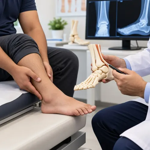
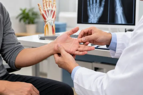
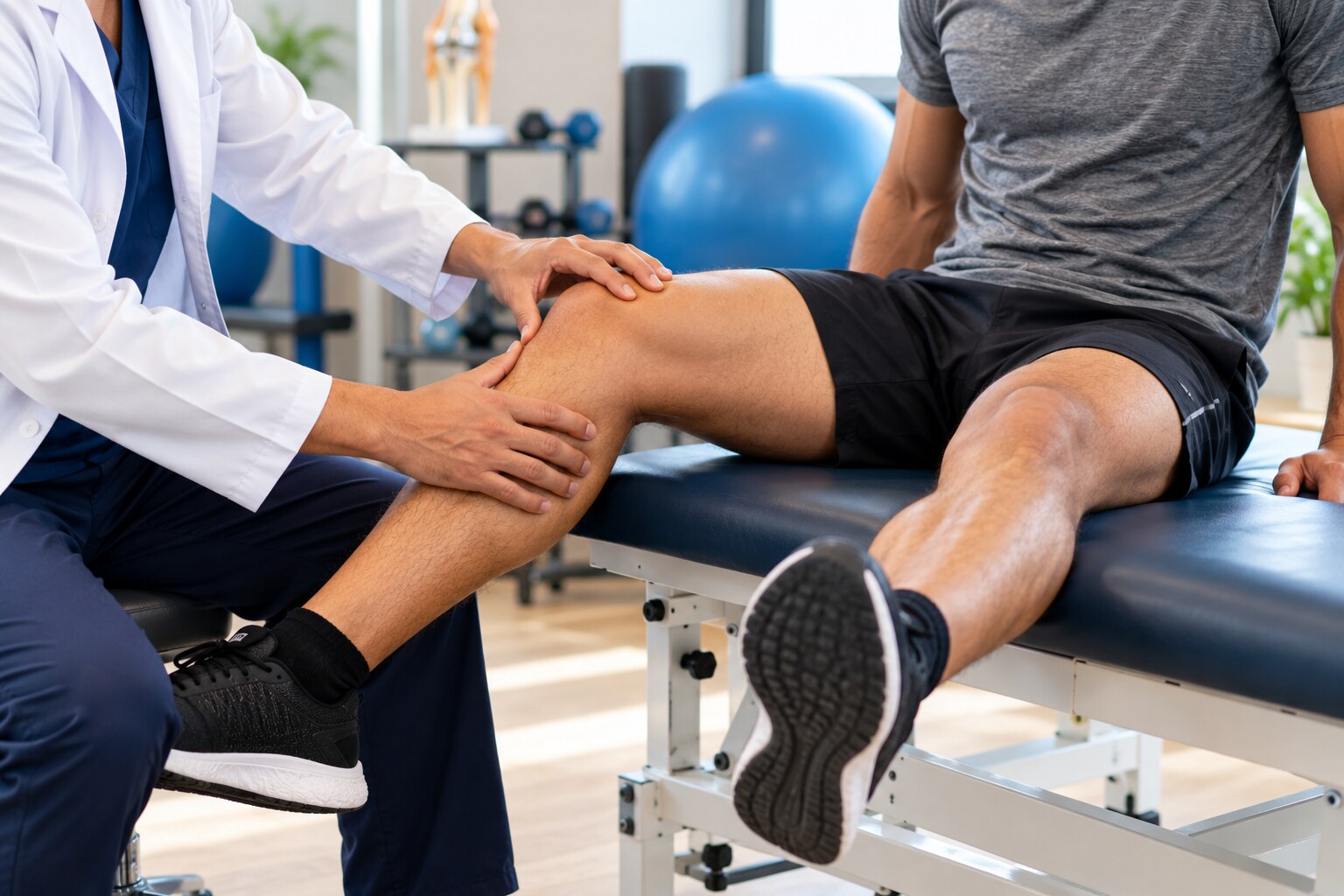
A sports medicine assessment starts with the injury mechanism, pain location, timing, swelling, bruising, instability and any change in movement. The clinician compares both sides, checks tenderness, range of motion, strength, joint stability, sensation, circulation and the ability to walk or perform a controlled movement.
For an ankle injury, the Ottawa Ankle Rules support radiographs when you have tenderness at the back edge or tip of either ankle bone, or cannot bear weight immediately and for four steps during assessment.
Foot X-rays are indicated for tenderness at the base of the fifth metatarsal or over the navicular, with the same weight-bearing test. These rules apply from age five and do not replace examination of deformity, open wounds or nerve and blood-vessel problems.
- A suspected concussion requires removal from play. Do not return on the same day; routine CT or MRI is unnecessary unless red flags suggest bleeding or another serious brain injury.
- Persistent focal pain, swelling or loss of function after a normal first scan requires reassessment, especially in children with possible growth-plate injury.
| Modality | Best use | Important limitation |
|---|---|---|
| X-ray | X-ray for fracture, dislocation or alignment problems | Can miss early stress fractures, cartilage damage and many ligament injuries |
| Ultrasound | Ultrasound tendon injury, muscle tears and fluid near the surface | Less useful for deep structures and some bone injuries |
| MRI | Early stress fracture, cartilage, ligament, tendon or muscle injury | Use it to answer a specific clinical question, not automatically after every injury |
Choosing Conservative Recovery, Rehabilitation or Surgery
Choose the least restrictive plan that protects the injured tissue while preserving safe movement. Conservative treatment suits many sprains, mild strains and tendinopathies; immobilisation or surgery becomes necessary when movement threatens healing, the joint is unstable, or tissue is displaced or completely torn.
| Plan | What it involves | When it applies |
|---|---|---|
| Conservative treatment | Relative rest, symptom control, progressive loading and physiotherapy rehabilitation | Stable sprains, muscle strains, tendinopathy and selected ACL injuries |
| Protection or immobilisation | Brace, boot, sling or protected weight bearing | Fractures, dislocations, severe sprains and selected bone stress injuries |
| Injection or surgery | A targeted injection, repair or reconstruction | Persistent inflammation, selected pain conditions, displaced fractures, ruptures or recurrent instability |
The injured structure determines the choice. A calf strain often improves with graded exercise, while a fracture or Achilles rupture needs protection and specialist care. Complete rest is not a definitive answer for Achilles tendinopathy: correct loading errors, rebuild calf strength and increase tendon load gradually.
Avoid corticosteroid injection around the Achilles because tendon weakening can increase rupture risk.
Bone stress injuries need load management, not pain suppression. Continued impact can turn a stress response into a fracture; high-risk sites such as the navicular, anterior tibial cortex and femoral neck need specialist assessment.
ACL rehabilitation may succeed without reconstruction when the knee is stable and goals are compatible. Recurrent giving-way, combined meniscal injury, pivoting-sport demands or failed rehabilitation can support surgery. Clearance requires strength, hop-test symmetry, movement quality, controlled swelling, psychological readiness and sport-specific progression—not time alone.
When to Get Specialist Care and How to Return to Sport
Self-care fits mild, improving pain without swelling, weakness, instability or altered movement. Choose physiotherapy when stiffness, weakness or altered mechanics persist; a sports physiotherapist can rebuild loading and sport-specific control. At Dhanvantari Hospital, ask whether your assessment needs sports medicine, an orthopaedic consultation or trauma care; urgency and instability determine the route.
| Option | What it means | When it applies |
|---|---|---|
| Self-care | Protect the area and reduce load | Symptoms steadily improve within a few days |
| Physiotherapy | Guided mobility, strength and movement retraining | Function remains limited after the acute phase |
| Sports medicine | Sport-specific assessment and graded loading | Pain recurs, performance drops or the diagnosis is unclear |
| Orthopaedic care | Assessment of bone, ligament, tendon or joint damage | Suspected fracture, dislocation, major ligament injury or failed rehabilitation |
| Trauma services | Immediate injury and surgical assessment | Open fracture, deformity, inability to bear weight, rapidly increasing swelling, numbness, cold or pale limb |
Return to sport requires more than pain relief. After ACL reconstruction, check full knee motion, controlled effusion, strength and hop-test symmetry, movement quality, psychological readiness and completion of sport-specific drills; time alone is not clearance.
- For concussion recovery, remove yourself immediately and do not return that day.
- Use relative rest for up to 48 hours, then progress through at least six stages, staying 24 hours at each stage.
- If symptoms return, step back to the last tolerated stage.
Related service
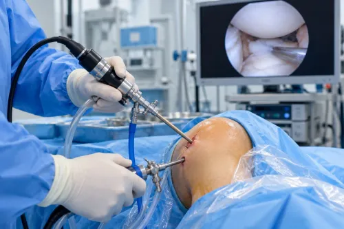
Arthroscopy and Sports Medicine Treatment Arthroscopy and Sports Medicine Treatment At Dhanvantari Hospital, Dr. Anand Upadhye and Dr. Sushant Upadhye provides advanced Arthroscopy and Sports Medicine treatments for patients... View service → |
Frequently asked questions
How can you tell normal soreness from an exercise-related injury?
Normal soreness is a dull, widespread ache that starts 12 to 24 hours after harder or unfamiliar activity, peaks within 24 to 72 hours and improves with movement. Injury is more likely when pain is sharp or localised, swelling or bruising appears, strength drops, or your walking changes.
What should you do during the first 24 to 48 hours after an exercise injury?
Stop the activity that caused pain, protect the injured area and use comfortable movement without forcing it. Seek prompt assessment for severe pain, deformity, inability to bear weight, marked swelling, numbness or worsening symptoms.
How do clinicians decide whether an exercise injury needs imaging?
Clinicians combine your injury mechanism, symptoms and physical examination. X-rays assess suspected fractures or dislocations; ultrasound can assess selected tendons and muscles; MRI provides detailed views of ligaments, cartilage, muscles and bone marrow.
When is surgery considered instead of conservative care or rehabilitation?
Conservative care and rehabilitation suit many muscle, tendon and joint injuries. Surgery enters the discussion when there is a displaced fracture, complete tendon or ligament tear, unstable joint, persistent functional loss or symptoms that do not improve with appropriate rehabilitation.
When can you return to sport after an exercise-related injury?
Return when daily movement and sport-specific drills are pain-free, swelling has settled, range of motion is restored and strength is comparable with the uninjured side. Increase training in stages rather than returning directly to full intensity.